This laboratory session is designed to introduce participants to the principles of microscopy through the examination of oral epithelium cells. The activity involves observing these cells in two conditions: their natural state with the addition of water and a stained state using Lugol’s solution to highlight the cell nuclei. This direct comparison aims to enhance the understanding of cellular morphology and the practical application of staining techniques in microscopy.
The main goal is to facilitate the microscopic observation of oral epithelium cells, emphasizing the differences between cells observed in their natural state and those where the nucleus is stained with Lugol’s solution.
Educational Goals
- Microscopy skills: Participants will learn how to properly use a microscope, focusing on the critical aspects of slide preparation and adjustment for clear observation.
- Cell morphology insight: This session aims to deepen the understanding of the structure of oral epithelium cells, enabling participants to distinguish cellular components under different conditions.
- Staining technique application: Introduces the concept and application of staining with Lugol’s solution, demonstrating its importance in enhancing the visibility of specific cell structures, such as the nucleus.
- Observation and documentation: Cultivates the ability to meticulously observe, accurately document, and interpret the microscopic details of cells, which are key skills in scientific research and reporting.
- Biological concepts application: Through practical experience, participants will apply theoretical knowledge of cell structure and function, reinforcing their learning through the direct observation of cells.
This laboratory session not only teaches the basics of microscopy and cell staining but also offers an invaluable hands-on experience. By observing oral epithelium cells under different conditions, participants will gain a comprehensive understanding of cell morphology, the significance of staining in biological observation, and the application of microscopy in the study of cellular structures.
Protocol
- Starting the microscope: Turn on the microscope by pressing the right switch (main light) onto the front of the device.
- Preparation of slides: Place two clean slides on your work surface.
- Sample application: Use the dropper to gently place 1 drop of buccal epithelium on each slide.
- Preparation of the first blade:
- Cover the slide with coverslip.
- Carefully blot the excess water with absorbent paper.
- Preparation of the second blade:
- Place a drop of Lugol on the second slide.
- Cover it with a coverslip.
- Blot the excess Lugol with absorbent paper.
- Observation of the first blade:
- Place the slide on the microscope stage.
- Click on the Microscope button on the tablet to view the microscope image.
- You can save an image of the observed microscope view by clicking the Save Image button located in the lower left area of the Microscope section.
- Microscope adjustment:
- You can adjust the magnification using the red screw on top of the microscope. Begin observation at a magnification of 40X.
- Fine-tune the focus using the coarse adjustment knobs located on each side of the microscope: green knob on the right (stage descends), blue knob on the left (stage rises).
- Gradually increase the magnification from 40X to 100X, then to 400X, adjusting the focus as required.
- Observation of the second blade:
- Replace the first slide with the second slide containing the cells and the Lugol’s iodine.
- Observe at 400X magnification to identify a cell nucleus, which should appear stained yellow by the Lugol.
- Recording of observations: Document or record important observations.
- Turning off the microscope: Turn off the device by deactivating the two switches at the front.
Anticipated Outcomes
Results are found in this document
In this lab, participants engage in a meticulous process to prepare and observe buccal epithelial cells under a microscope. This exercise not only enhances understanding of cell structure but also hones laboratory skills.
- Cell visualization: Participants will successfully prepare slides with buccal epithelial cells and use a microscope to observe these cells at various magnifications. The water-prepared slide will provide a clear view of the cells’ general structure, while the iodine-stained slide will highlight specific cell components, like the nucleus, making them more distinguishable.
- Staining effectiveness: The iodine solution will stain the cell components, particularly the nucleus, enabling observers to note the distinct parts of the cell. This contrast is crucial for understanding cell compartmentalization and function.
- Microscopy skills: Through adjusting the microscope’s focus and changing magnifications, participants will gain practical experience in using this essential scientific tool, learning to identify and record significant cellular details.
Significance and Lessons Learned:
- Cellular biology insights: Observing buccal epithelial cells provides a concrete understanding of cell theory, illustrating the cell’s structural and functional units. This hands-on experience cements theoretical knowledge with practical observation.
- Technical proficiency: Mastery in preparing slides, handling delicate laboratory equipment, and conducting precise observations are key skills developed in this lab. These are fundamental in various scientific investigations, where careful preparation and acute observational abilities are paramount.
- Analytical skills: Interpreting the observed cellular structures fosters analytical thinking, as participants correlate cell morphology with function, enhancing their understanding of biological processes at the microscopic level.
- Safety and protocol adherence: The emphasis on safety measures and protocol adherence instills a sense of responsibility and rigor, essential attributes for any scientific endeavor.
Ultimately, this laboratory exercise is not just about observing cells; it’s an integrated learning experience that develops a range of skills and deepens understanding of fundamental biological principles. Participants leave with a greater appreciation for the microscopic world and its relevance to broader biological contexts.
Summary of Assignment by Grade Range
Grades 3-5 (Ages 8-10)
- Focus: Basic introduction to microscopy and simple cell observations.
- Activities: Using microscopes, preparing simple slides, observing natural and stained cells, basic safety instructions.
Grades 6-8 (Ages 11-13)
- Focus: Intermediate skills in microscopy and understanding cell morphology.
- Activities: Preparing and staining slides, using microscopes, observing and documenting cell morphology, following detailed safety protocols.
Grades 9-12 (Ages 14-18)
- Focus: Advanced microscopy skills and in-depth analysis of cell structure.
- Activities: Mastering microscope use, preparing and staining slides, detailed observation and analysis, meticulous documentation, adhering to advanced safety protocols.
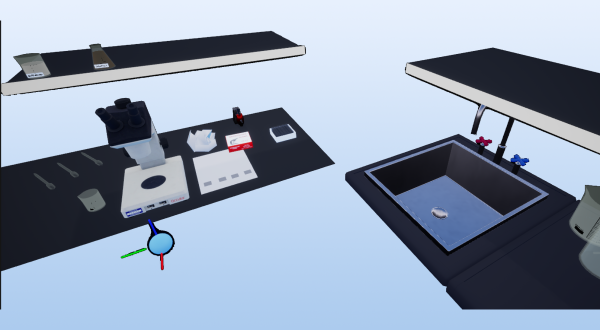
Laboratory essentials
Instruments
- Beaker (50 ml)
- Droppers
- Microscope
- Microscope blades
- Microscope sliders
- Paper towel
- Tweezers
Products
- Human buccal epithelium cells in suspension
- Lugol 2% solution